๐Ÿพ Veterinary Imaging

Expert reporting for
every scan you take โ€” fast.

CT, MRI, radiography, ultrasound. Submit your DICOM files and receive a specialist-written report โ€” board-certified, personally reviewed, delivered within 24-48 hours.

4Imaging modalities
24โ€“48hrReport turnaround
100%Personally reviewed

Choose a modality below

โ˜ข๏ธ
CT Reporting
Thorax, abdomen, orthopaedics
๐Ÿงฒ
MRI Reporting
Neurology, brain, spinal cord, MSK
๐Ÿฉป
Radiography
Thorax, abdomen, orthopaedic
๐Ÿ”Š
Ultrasound
Abdominal, cardiac, interventional

CT reporting that
can catch what others miss.

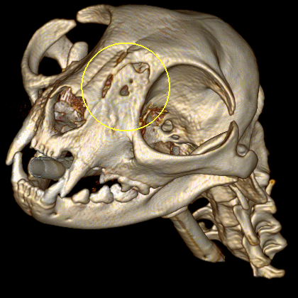
Computed tomography is one of the most powerful diagnostic tools in veterinary medicine โ€” but only when read by someone who does it every day. ImagingVets has interpreted thousands of CT studies across thorax, abdomen, and musculoskeletal cases. You get a report that tells you exactly what's there and what it means.


Typical turnaround: Next business day standard.


Submit CT Case โ†’
3D CT reconstruction of a cat skull
CT โ€” Veterinary

What we report on.

Thoracic Masses

Primary lung tumours, metastatic disease, mediastinal masses, and pleural abnormalities.

Portosystemic Shunts

Intrahepatic and extrahepatic shunts โ€” full vascular mapping and surgical planning support.

Oncology Staging

Full body staging for lymphoma, carcinomas, and sarcomas with detailed nodal assessment.

Orthopaedic Pathology

Elbow dysplasia, OCD, fragmented coronoid, joint disease, and complex fracture planning.

Neck & Cervical Spine

Thyroid masses, oesophageal abnormalities, cervical disc disease, and vascular anomalies.

Nasal & Sinus Disease

Nasal tumours, rhinitis, fungal disease, and dental/jaw pathology requiring CT for characterisation.

From scan to report.

01

Acquire DICOM Files

Perform the CT at your own facility. Export DICOM files from your scanner as usual.

02

Submit Online

Upload via our secure portal with patient details and your clinical question. Takes under 5 minutes.

03

Radiologist Reviews

Every CT is reviewed by board certified radiologists.

04

Report Delivered

You receive a structured, clinician-ready report addressing your specific query.

Ready to submit your CT case?

Secure upload ยท 24โ€“48hr turnaround

Neurological cases
need expert eyes.

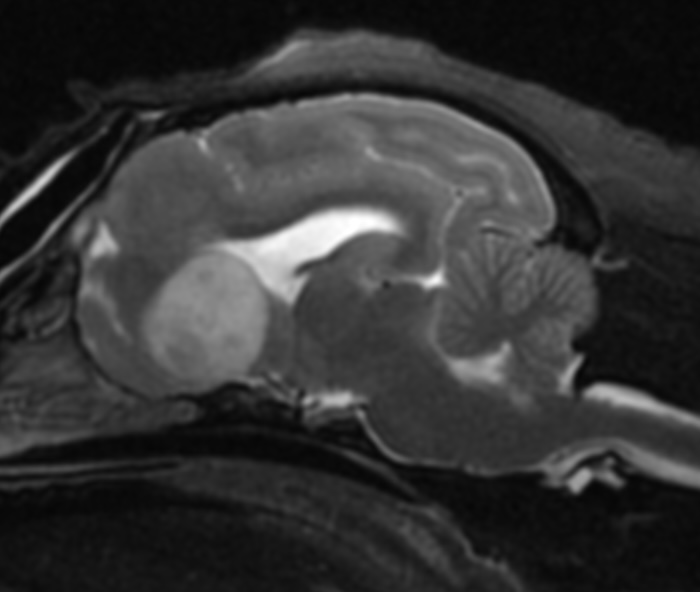
MRI is the gold standard for brain and spinal cord disease โ€” and the most technically demanding modality to interpret. Dr Hoffmann reports for specialist neurology groups to give clear, differential-driven reporting that directly informs their clinical decision-making, from disc disease to tumour characterisation. MRI is also used to help in the assessment of MSK diseases.


Typical turnaround: Next business day standard.


Submit MRI Case โ†’
MRI study of a brain
MRI โ€” Veterinary

What we report on.

Intracranial Disease

Brain tumours, encephalitis, hydrocephalus, vascular events โ€” characterised and graded.

Intervertebral Disc Disease

Hansen Type I & II, disc extrusion vs. protrusion, cord compression grading.

Spinal Cord Tumours

Intramedullary, intradural-extramedullary, and extradural mass localisation and characterisation.

Vestibular Disease

Central vs peripheral differentiation, inner ear pathology, brainstem lesions.

Soft Tissue Masses

Musculoskeletal tumours, infiltrative lesions, and perineural sheath tumours.

Lumbosacral Disease

Degenerative lumbosacral stenosis, cauda equina syndrome, foraminal narrowing.

From scan to report.

01

Acquire DICOM Files

Complete your MRI sequences at your facility. Export all sequences as DICOM.

02

Submit + Clinical History

Upload with a brief clinical history and your specific question for the most targeted report.

03

Expert MRI Review

Dr Hoffmann reviews all sequences and constructs a differential-driven assessment.

04

Structured Report

Findings, interpretation, differentials and recommendations โ€” delivered to your inbox.

Neurological case needs reading?

24โ€“48hr turnaround ยท Differential-driven reports ยท Personally reviewed

A second opinion on
your radiographs.

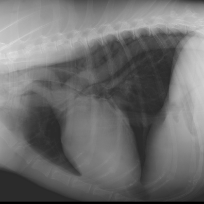
Radiography is the most commonly used diagnostic imaging modality in practice โ€” and the one where important findings are often missed under time pressure in a busy practice. A specialist radiograph report can change your diagnosis, change your treatment, and protect your patient.


Submit Radiographs โ†’
Thoracic radiograph
Radiography โ€” Veterinary

What we report on.

Thoracic Radiographs

Cardiac silhouette, pulmonary patterns, pleural space, and mediastinal assessment.

Abdominal Radiographs

GI obstruction, organ size assessment, mass effect, peritoneal fluid, and mineralisation.

Orthopaedic Radiographs

Fractures, luxations, joint disease, and developmental conditions in growing animals.

Dental Radiographs

Tooth root disease, periapical lesions, resorptive disease, and jaw pathology.

Exotic Pets

Avian, reptile, and small mammal radiographs interpreted with species-specific expertise.

Whole Body Studies

Metastasis checks, trauma surveys, and multi-region assessments in a single report.

From X-ray to expert report โ€” same day.

01

Take Your Radiographs

Use your existing digital X-ray system. All standard digital formats accepted.

02

Submit in Minutes

Upload files with patient signalment and your clinical question via the secure portal.

03

Specialist Review

Dr Hoffmann reviews every view, noting incidental findings as well as primary concerns.

04

Report in Your Inbox

A clear, systematic report ready to share with your client or referring specialist.

Don't sit on an uncertain radiograph.

Fast turnaround ยท Clear language ยท Actionable recommendations

Advanced ultrasound,
at your clinic.

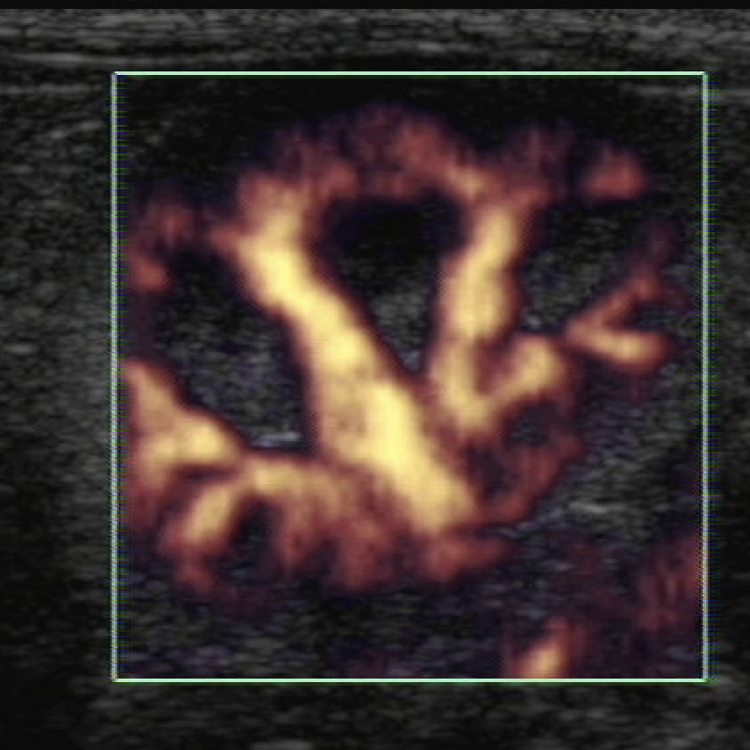
Ultrasound is operator-dependent โ€” which means results vary enormously. Dr Hoffmann can both report on ultrasound images submitted remotely and visit your clinic to perform advanced studies you can't do yourself. Complex abdominal, screening echocardiography, and interventional โ€” all on-site.


Options: Submit images remotely for reporting, or book an on-site visit.


Submit Ultrasound Images โ†’ Request On-Site Visit
Colour Doppler ultrasound study
Ultrasound โ€” Veterinary

What we cover.

Echocardiography

Full cardiac function assessment, valve disease, cardiomyopathy, congenital defects.

Abdominal Ultrasound

Liver, spleen, kidneys, adrenals, GI tract, bladder, reproductive organs โ€” complete survey.

Mass Characterisation

Distinguishing benign from suspicious masses; guiding FNA and biopsy decisions.

Free Fluid Assessment

FAST protocol, effusion characterisation, guided drainage and diagnostic sampling.

Musculoskeletal

Tendon and ligament injuries, joint effusion, soft tissue swellings.

Interventional Ultrasound

Ultrasound-guided FNA, biopsy, cystocentesis, and fluid sampling performed on-site.

How a clinic visit works.

01

Request a Visit

Submit your ultrasound enquiry โ€” include the patient details and study required.

02

Confirm Booking

We'll confirm availability and visit date. You prepare the patient as normal.

03

Dr Hoffmann Attends

She brings her own equipment and performs the full study at your clinic.

04

Report + Discussion

Findings discussed with you on the day, with written report to follow.

Need advanced ultrasound at your clinic?

Remote reporting or on-site visits โ€” whichever your case needs